|
Scoring of tissue microarray images
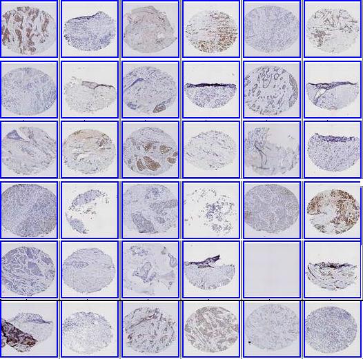
The tissue microarray (TMA) image measures tumor-specific protein expression level of the donor's block. It has emerged as an important high-throughput tool for the evaluation of histology-based laboratory tests. It is used extensively in cancer studies, including clinical outcome analysis, tumor progression analysis, the identification of diagnostic or prognostic factors etc, and the development and validation of tumor-specific biomarkers. The readings of a TMA image are quantified by its staining pattern, which is typically summarized as a numerical score by the pathologist. Our work aims at reducing the inherent variability and subjectivity with manual scoring, and to report scores in real-time.
1. TACOMA
-- A tissue image scoring algorithm that makes use of statistical regularity in the tissue images, incorporates pathologists' knowledge in the form of representative image patches. The algorithm can detect salient pixels that would be used by pathologists in their reading of TMA images.
2. deepTacoma
-- Extension of the TACOMA algorithm that leverages the latent clustering structures in the data, and turn such information into easily deployable regularizing features.
|
|
Citation
[1] D. Yan, P. Wang, B. S. Knudsen, M. Linden and T. W. Randolph.
Statistical methods for tissue array images--algorithmic scoring and co-training.
Annals of Applied Statistics, Vol 6(3), 1280-1305, 2012.
arXiv:1102.0059
[2] D. Yan, T. W. Randolph, J. Zou and P. Gong.
Incorporating deep features in the analysis of tissue array images.
Statistics and its Interface, Vol 12(2), 283-293, 2019.
arXiv:1812.00887.
[3] D. Yan, J. Zou and Z. Li. Learning low-dimensional manifolds for scoring of tissue microarray
images. arXiv:2102.11396